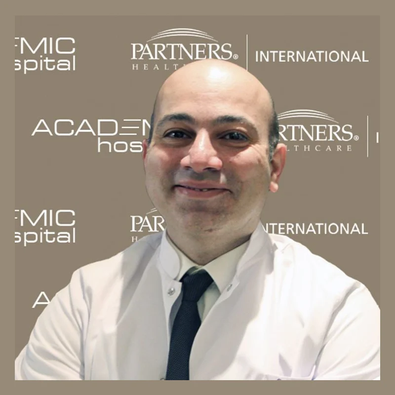
Dr. Öğr. Üyesi Görkem Kıyak
OrthopedicsVerified
- 21+years exp.
English · Turkish
FromPrice on request
Free quote
60 seconds · no obligationView Profile
Valgus deformity is an orthopaedic condition where a bone or joint segment is abnormally angled outward away from the body's midline, commonly seen at the knees or big toes. Individuals seek medical evaluation due to persistent joint pain, difficulty walking, joint instability, or visible structural changes. Available corrective treatment options depend on a detailed specialist assessment of alignment, joint wear, and patient mobility. Compare hospitals in Turkey that treat Valgus Deformity and get a free review of your medical records.

1 doctor Compare doctors, ratings and prices — ask any of them for a free quote.
OrthopedicsVerified
English · Turkish
60 seconds · no obligationView Profile
Valgus deformity is an orthopaedic condition where a bone or joint segment is abnormally angled outward away from the body's midline, commonly seen at the knees or big toes. Individuals seek medical evaluation due to persistent joint pain, difficulty walking, joint instability, or visible structural changes. Available corrective treatment options depend on a detailed specialist assessment of alignment, joint wear, and patient mobility.
Specialty: Orthopedics
Depending on the stage and your medical history, doctors may recommend:
The cost of treating Valgus Deformity depends on the diagnosis and the treatment chosen. Doctors first review your medical records — free of charge — and you then receive an itemised quote.
Options may include Surgical Treatment of Valgus Deformity, Hallux Valgus Surgery, Lapiplasty 3D Bunion Correction, Hallux Valgus 3D Lapiplasty. Your doctor chooses the approach after reviewing your tests and scans.
Valgus Deformity is treated by Orthopedics specialists. 2 clinics in Turkey on our platform offer this treatment.
A valgus deformity is an orthopaedic structural misalignment where a bone or joint segment angles outward away from the body's central midline. The most common clinical manifestations occur at the knee (knock-knees or genu valgum) and the first metatarsal joint of the foot (hallux valgus, frequently referred to as a bunion). It can also affect the hip (coxa valga) or elbow (cubitus valgus). When joint geometry is altered, physical stress is distributed unevenly across articular cartilage, joint capsules, and supporting ligaments.
Patients experiencing valgus alignment often report a distinct set of progressive symptoms, including:
Accurate correction of a valgus alignment requires precise imaging and physical assessment. Specialist centers providing Orthopedics care in Turkey follow standardized diagnostic pathways to map the exact angular deviation before recommending an intervention.
Upon arrival, your clinical team will perform a physical examination to test joint stability, gait mechanics, and range of motion. Diagnostic imaging typically includes:
To streamline your evaluation, bring previous weight-bearing X-rays, MRI reports, gait analysis documentation, and a summary of past non-surgical treatments (such as custom orthotics or physical therapy records).
Management ranges from conservative interventions to structural surgical realignment. Treatment selection depends on patient age, bone maturity, severity of the angle, and the condition of the underlying cartilage.
| Option | Typically used when | Things to know |
|---|---|---|
| Conservative Management | Mild angular deviation, minimal functional impairment, or flexible deformities. | Includes custom orthotic insoles, foot realignment splints, targeted physical therapy, and activity modification. Manages symptoms but does not alter fixed bone geometry. |
| Realignment Osteotomy | Moderate to severe deformities in younger or active patients with preserved joint space. | The surgeon cuts and realigns the bone (such as a distal femoral osteotomy for knee valgus or a scarf/chevron osteotomy for hallux valgus) using plates or screws. Preserves the natural joint. |
| Joint Replacement Surgery | Advanced valgus deformity combined with severe end-stage joint degeneration or arthritis. | Replaces damaged joint surfaces with prosthetic components while correcting the overall mechanical axis. Common for severe knee or hip deformities. |
Depending on overall physical health and medical history, some patients undergoing orthopaedic evaluation may also discuss broader health assessments, such as Check-ups & Diagnostics or specialized medical support via Rehabilitation services.
Traveling abroad for bone realignment requires careful timing. The overall itinerary depends on whether your treatment is non-surgical or invasive.
Before traveling, send recent X-rays and clinical histories to the medical team in Turkey. Specialists review your mechanical axis measurements to provide an initial assessment of feasibility, potential surgical techniques, and expected recovery timelines.
If undergoing a surgical realignment or osteotomy, patients usually remain in hospital for 2 to 4 days for pain management and early mobilization. You should plan to stay in Turkey for 10 to 14 days post-discharge. This allows for wound monitoring, suture removal, initial physical therapy sessions, and a final fit-to-fly assessment by your treating surgeon.
Bone healing takes several months. Before departing Turkey, you will receive your operative notes, discharge summary, and digital imaging copies. You must arrange post-operative follow-up with a local orthopaedic physician or physical therapist in your home country to guide your ongoing rehabilitation and weight-bearing progression.
Long-distance travel introduces specific medical considerations, especially following lower-limb orthopaedic procedures. Air travel increases the risk of deep vein thrombosis (DVT). Patients who have recently undergone osteotomy or joint replacement must follow prescribed anti-coagulation protocols and wear compression garments as advised by their clinical team.
Immediate travel is contraindicated if you have an active systemic infection, unstable cardiovascular conditions, or acute soft-tissue compromise around the target joint. Surgical intervention should be delayed until these conditions are fully managed.
Whether in your hotel in Turkey or after returning home, contact emergency medical care immediately if you notice:
All surgical decisions, weight-bearing schedules, and travel permissions are determined by the treating surgeon following a comprehensive direct examination and review of imaging findings.
Valgus deformities can be congenital, developing during growth due to genetic factors or developmental bone variations. They can also develop later in life secondary to joint trauma, asymmetrical growth plate injuries, structural ligament laxity, metabolic bone disorders, or progressive mechanical wear associated with osteoarthritis.
No, surgery is not always required. Mild deformities causing minimal discomfort are often managed conservatively with custom orthotics, targeted physical therapy, and activity modification. Surgical realignment is generally reserved for patients experiencing severe pain, significant functional impairment, progressive joint wear, or structural instability that fails to improve with conservative care.
Initial bone healing typically takes 6 to 8 weeks, during which weight-bearing may be restricted using crutches or specialized braces. Full recovery, including complete bone consolidation, muscle strength restoration, and return to high-impact activities, often takes between 6 and 12 months, guided by structured physical therapy.
While modern surgical techniques aim to permanently alter the mechanical axis, partial recurrence is possible in rare cases. Factors influencing long-term outcomes include bone quality, compliance with post-operative weight-bearing guidelines, ligament balance, and ongoing joint wear. Your surgeon will discuss specific long-term expectations during your consultation.
Most orthopaedic surgeons recommend waiting at least 10 to 14 days after surgical realignment before taking a flight. This timeframe allows initial wound healing, reduces the acute risk of deep vein thrombosis, and ensures a final physical check-up and fit-to-fly clearance can be performed before departure.
Your request is in!
Your coordinator will contact you — usually within 2 hours, 24/7.
Want a faster, more accurate assessment?
Add photos & medical detailsLeave your number — a coordinator calls you in your language, usually within 15 minutes during office hours.
Thank you!
A coordinator will contact you at the time you chose.

