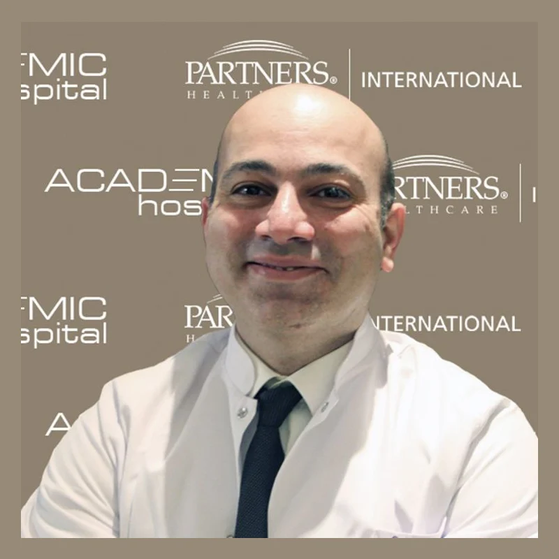
Dr. Öğr. Üyesi Görkem Kıyak
OrthopedicsVerified
- 21+years exp.
English · Turkish
FromPrice on request
Free quote
60 seconds · no obligationView Profile
Osteochondrosis refers to a group of conditions that disrupt bone growth and cartilage development at the joint surfaces or growth plates. It frequently affects growing children and active adolescents, who often present with persistent joint pain, swelling, tenderness, and restricted mobility. Specific treatment plans vary widely and depend upon a thorough specialist assessment of skeletal maturity and severity. Compare hospitals in Turkey that treat Osteochondrosis and get a free review of your medical records.

1 doctor Compare doctors, ratings and prices — ask any of them for a free quote.
OrthopedicsVerified
English · Turkish
60 seconds · no obligationView Profile
Osteochondrosis refers to a group of conditions that disrupt bone growth and cartilage development at the joint surfaces or growth plates. It frequently affects growing children and active adolescents, who often present with persistent joint pain, swelling, tenderness, and restricted mobility. Specific treatment plans vary widely and depend upon a thorough specialist assessment of skeletal maturity and severity.
Specialty: Orthopedics
The cost of treating Osteochondrosis depends on the diagnosis and the treatment chosen. Doctors first review your medical records — free of charge — and you then receive an itemised quote.
Osteochondrosis is treated by Orthopedics specialists. 2 clinics in Turkey on our platform offer this treatment.
Osteochondrosis describes a group of developmental disorders that disrupt the normal blood supply to growing bones and cartilage. This interruption affects the growth plates (apophyses or epiphyses) and joint surfaces, leading to localised tissue necrosis, cartilage fragmentation, and secondary inflammation. While the condition commonly affects growing children and active adolescents during growth spurts, certain forms can also impact adults.
Depending on which joint or growth plate is involved, osteochondrosis presents under specific clinical names. Common forms include Legg-Calvé-Perthes disease (hip joint), Osgood-Schlatter disease (tibial tuberosity below the knee), Scheuermann's disease (thoracic or lumbar spine), and Freiberg's disease (metatarsal head in the foot). Regardless of the specific site, the primary mechanism involves temporary vascular insufficiency at the ossification centre.
Patients seeking care through Orthopedics specialists typically present with a predictable set of symptoms:
An accurate diagnosis requires a thorough physical evaluation combined with targeted diagnostic imaging to establish the stage and severity of the bone changes. When travelling for specialized evaluation, international patients complete a comprehensive clinical assessment to differentiate osteochondrosis from infection, inflammatory arthritis, or acute traumatic fractures.
Diagnostic pathways at an overseas medical centre typically involve:
Patients travelling abroad should bring previous X-rays, MRI scans (on DICOM disc or digital format), medical reports detailing the onset of symptoms, and records of any previous conservative treatments or surgical interventions. Comprehensive Check-ups & Diagnostics help the local clinical team confirm the exact stage of skeletal maturity before finalising a care plan.
Management depends heavily on the patient's skeletal age, the precise anatomical site, and whether the joint surface remains intact. Most childhood and adolescent cases respond well to conservative measures, while persistent, unstable, or adult forms may require surgical intervention.
| Option | Typically used when | Things to know |
|---|---|---|
| Conservative Management | Early-stage disease, stable growth plate lesions, and skeletally immature patients. | Focuses on activity modification, targeted immobilization, pain management, and specialized physical therapy. |
| Arthroscopic Debridement | Unstable cartilage fragments, loose bodies in the joint space, or persistent pain. | Minimally invasive procedure to smooth damaged surfaces and remove free fragments; frequently coordinated alongside Rehabilitation services. |
| Surgical Fixation or Grafting | Advanced osteochondritis dissecans with large, detached fragments or structural joint collapse. | Requires securing the fragment or performing bone grafting to re-establish joint surface stability. |
In pediatric patients, treatment aims to preserve joint congruity while the bone revascularises naturally over time. Medical decisions are made strictly by the treating orthopaedic surgeon following direct examination and review of up-to-date imaging.
International patients planning care in Turkey should begin with a remote evaluation of their diagnostic files. Turkish medical facilities review imported MRI discs and medical histories to outline a preliminary pathway before travel. Patients undergoing non-surgical management typically spend several days in-country for specialist consultations, custom bracing, and physical therapy instruction. If surgery is required, stays generally extend from one to two weeks to allow for immediate postoperative monitoring and initial mobility training.
Before flying home, patients receive detailed guidance on weight-bearing restrictions, wound care, and structured exercise regimens. Follow-up care is maintained through remote consultations with the Turkish clinical team in coordination with the patient's local general practitioner or orthopaedic specialist at home.
Long-distance travel is contraindicated if a patient has an acute, unstable joint condition, severe unmanaged pain, or a recent postoperative wound that has not healed sufficiently. Sitting for long flights with acute lower limb or spinal inflammation can exacerbate pain and increase the risk of deep vein thrombosis.
Patients must seek immediate local emergency care rather than attempting long-distance travel if they develop any of the following warning signs:
Osteochondrosis develops when the blood supply to a bone's growth plate or joint surface is temporarily disrupted. The exact cause is often a combination of micro-trauma, repetitive mechanical stress during physical growth, genetic predisposition, and localized vascular variations. It is most frequently observed in active children and adolescents during periods of rapid bone growth.
Recovery timelines vary widely depending on the patient's skeletal maturity, the specific joint affected, and the severity of the lesion. Conservative management in growing children may require several months to a year for full bone remodeling. Post-surgical recovery generally involves six to twelve weeks of restricted weight-bearing, followed by structured physical therapy over several months.
While osteochondrosis primarily affects growing children and adolescents, adults can suffer from the late effects of childhood osteochondrosis or develop adult-onset osteochondritis dissecans. In adults, the condition typically involves persistent joint surface irregularities or loose cartilage fragments, which often require surgical evaluation to prevent premature joint degeneration.
You should collect all recent diagnostic imaging, particularly high-resolution MRI and X-ray files stored on a CD or digital cloud folder. Include official radiologist reports, notes from previous orthopaedic consultations, details of prior physical therapy, and a list of current medications. Bringing complete records ensures the consulting team in Turkey can accurately evaluate your condition.
Your treating surgical and orthopaedic team in Turkey will provide a comprehensive, structured rehabilitation plan upon discharge. You will transfer this plan to a local physiotherapist in your home country. Remote consultations between your local therapist and the international hospital team can be arranged to monitor your progress safely.
Your request is in!
Your coordinator will contact you — usually within 2 hours, 24/7.
Want a faster, more accurate assessment?
Add photos & medical detailsLeave your number — a coordinator calls you in your language, usually within 15 minutes during office hours.
Thank you!
A coordinator will contact you at the time you chose.

