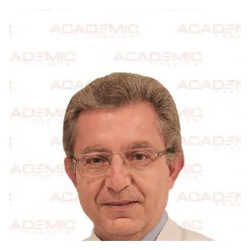
Prof. Dr. Ali Serdar Fak
Cardiac SurgeryVerified
- 29+years exp.
English · Turkish
FromPrice on request
Free quote
60 seconds · no obligationView Profile
A ventricular septal defect is a congenital heart condition characterised by an opening in the septum separating the lower heart chambers. It affects infants and can persist into adulthood, causing breathlessness, poor feeding, or fatigue that prompts medical consultation. Necessary monitoring or corrective intervention options depend on a thorough clinical assessment by a cardiology specialist. Compare hospitals in Turkey that treat Ventricular Septal Defect and get a free review of your medical records.

2 doctors Compare doctors, ratings and prices — ask any of them for a free quote.
Cardiac SurgeryVerified
English · Turkish
60 seconds · no obligationView Profile
Cardiac SurgeryVerified
English · Turkish
60 seconds · no obligationView Profile
A ventricular septal defect is a congenital heart condition characterised by an opening in the septum separating the lower heart chambers. It affects infants and can persist into adulthood, causing breathlessness, poor feeding, or fatigue that prompts medical consultation. Necessary monitoring or corrective intervention options depend on a thorough clinical assessment by a cardiology specialist.
Specialty: Cardiac Surgery
Depending on the stage and your medical history, doctors may recommend:
The cost of treating Ventricular Septal Defect depends on the diagnosis and the treatment chosen. Doctors first review your medical records — free of charge — and you then receive an itemised quote.
Options may include Ventricular Septal Defect Surgery, Interventricular Septal Defect Surgery. Your doctor chooses the approach after reviewing your tests and scans.
Ventricular Septal Defect is treated by Cardiac Surgery specialists. 2 clinics in Turkey on our platform offer this treatment.
A Ventricular Septal Defect (VSD) is a structural birth anomaly characterized by an abnormal opening in the interventricular septum, the muscular wall separating the lower chambers of the heart. This opening allows oxygen-rich blood from the left ventricle to mix with oxygen-poor blood in the right ventricle, increasing the total volume of blood pumped towards the lungs. VSD is often identified during early childhood, but smaller or moderate defects can remain undetected until adolescence or adulthood.
The clinical presentation varies depending on the size and location of the hole within the septum. Infants with moderate to large defects typically display signs of heart failure within the first few weeks of life, including rapid breathing, excessive sweating during feeds, poor weight gain, and recurrent chest infections. In older children and adults, symptoms may manifest as persistent fatigue, decreased exercise tolerance, heart palpitations, or shortness of breath. Over time, an uncorrected large VSD can lead to pulmonary arterial hypertension, heart failure, and an increased risk of infective endocarditis.
Initial evaluation often begins when a physician detects a characteristic holosystolic heart murmur during a physical examination. To confirm the diagnosis and assess the defect's hemodynamic impact, specialist centers in Turkey perform comprehensive non-invasive and invasive diagnostic tests. Patients traveling for specialized evaluation are advised to compile their medical records beforehand, including previous clinical notes, medication lists, and prior diagnostic imaging.
Key diagnostic investigations usually involve:
These procedures are frequently coordinated alongside initial evaluations within specialized departments like Cardiac Surgery or Paediatrics to establish an accurate surgical plan.
Management strategies range from conservative clinical observation to active interventional closure. Small VSDs that do not cause heart strain or pulmonary hypertension often close spontaneously during early childhood or require no intervention other than routine monitoring. However, intervention is required when the defect causes symptomatic heart failure, significant left heart enlargement, or elevated pulmonary artery pressures. The appropriate approach is decided by the treating multidisciplinary team following direct review of diagnostic findings.
| Option | Typically used when | Things to know |
|---|---|---|
| Clinical Monitoring | Small, asymptomatic VSDs with normal pulmonary artery pressures. | Requires periodic echocardiograms; spontaneous closure can occur in early childhood. |
| Transcatheter Device Closure | Muscular or selected perimembranous VSDs with suitable anatomical margins. | Minimally invasive approach via the femoral vein; avoids open-heart surgery and shortens recovery time. |
| Surgical Patch Repair | Large VSDs, perimembranous or outlet defects near heart valves, or failed catheter closures. | Performed via open-heart surgery using a synthetic patch or tissue graft under cardiopulmonary bypass. |
In complex congenital cases where additional structural repairs or diagnostics are necessary, multidisciplinary teams may involve specialists from Pulmonology & Thoracic Surgery or Check-ups & Diagnostics to manage overall heart and lung function.
International travel for cardiac care requires thorough advanced planning. Before traveling, patients or their guardians submit detailed medical records, echocardiograms, and relevant test results for preliminary remote review. Once accepted, the clinical team coordinates direct evaluations upon arrival to confirm suitability for catheter-based or open surgical intervention.
The length of stay varies significantly by the chosen procedure. Transcatheter closures typically require a stay of 3 to 5 days, including 1 to 2 days in the hospital for observation. Surgical patch repairs require a longer stay, usually 10 to 14 days in Turkey, which includes time in an intensive care unit, step-down ward care, and outpatient checks prior to flying home. Before discharge, the surgical team provides clear guidance regarding post-procedure medication, activity restrictions, wound care, and recommendations for long-term follow-up with a local cardiologist in the patient's home country.
Patients with severe symptomatic VSD, severe pulmonary arterial hypertension (Eisenmenger syndrome), or severe uncontrolled heart failure must avoid air travel until thoroughly evaluated and stabilized by a cardiologist. Rapid atmospheric pressure changes can exacerbate hypoxia and right-to-left shunting in advanced cases.
Immediate local medical care is required if a patient experiences any of the following warning signs before or after treatment:
A Ventricular Septal Defect is a congenital heart condition, meaning it develops before birth while the baby's heart is forming. It occurs when the muscular wall separating the lower chambers does not close completely. While most cases occur spontaneously without a clear cause, genetic factors and environmental influences during pregnancy can contribute to its development.
Yes, small Ventricular Septal Defects, particularly those located in the muscular part of the septum, frequently close on their own during the first few years of life. However, larger defects or those situated near heart valves rarely close spontaneously and usually require medical or surgical intervention to prevent long-term complications.
The required stay depends on the procedure type. Minimally invasive transcatheter closure generally requires 3 to 5 days in the country. Open surgical repair requires approximately 10 to 14 days to allow for hospital recovery, post-operative monitoring, and final medical clearance before flying.
Device closure is a catheter-based technique where a small closure device is guided to the heart through a blood vessel in the groin. Open repair involves traditional surgery under cardiopulmonary bypass to place a patch over the hole. The choice depends on the size, exact location, and structure of the defect.
Air travel safety depends on individual recovery and procedure type. Patients who undergo transcatheter closure can often fly within 1 to 2 weeks, whereas open surgery patients usually require 3 to 4 weeks before traveling by air. Final clearance must always be provided by the treating cardiologist.
Your request is in!
Your coordinator will contact you — usually within 2 hours, 24/7.
Want a faster, more accurate assessment?
Add photos & medical detailsLeave your number — a coordinator calls you in your language, usually within 15 minutes during office hours.
Thank you!
A coordinator will contact you at the time you chose.