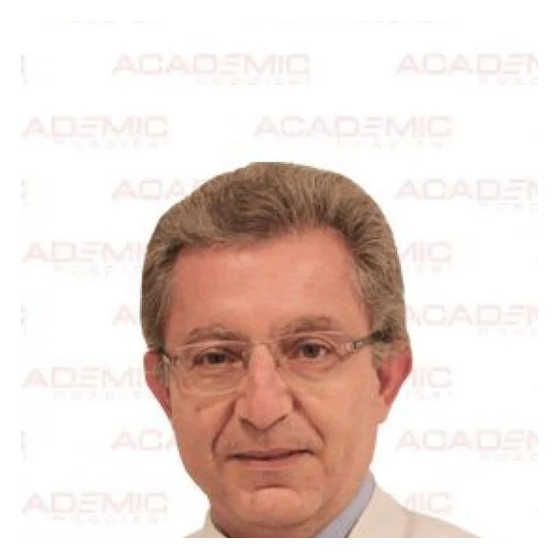
Prof. Dr. Ali Serdar Fak
Cardiac SurgeryVerified
- 29+years exp.
English · Turkish
FromPrice on request
Free quote
60 seconds · no obligationView Profile
Aortic valve insufficiency is a cardiac condition in which the aortic valve fails to close tightly, permitting blood to leak backward into the left ventricle. It can affect adults due to valve degeneration, hypertension, or infections. People consult medical professionals because of fatigue, exertional breathlessness, palpitations, or light-headedness. Tailored treatment options depend on an extensive specialist assessment. Compare hospitals in Turkey that treat Aortic Valve Insufficiency and get a free review of your medical records.

2 doctors Compare doctors, ratings and prices — ask any of them for a free quote.
Cardiac SurgeryVerified
English · Turkish
60 seconds · no obligationView Profile
Cardiac SurgeryVerified
English · Turkish
60 seconds · no obligationView Profile
Aortic valve insufficiency is a cardiac condition in which the aortic valve fails to close tightly, permitting blood to leak backward into the left ventricle. It can affect adults due to valve degeneration, hypertension, or infections. People consult medical professionals because of fatigue, exertional breathlessness, palpitations, or light-headedness. Tailored treatment options depend on an extensive specialist assessment.
Specialty: Cardiac Surgery
The cost of treating Aortic Valve Insufficiency depends on the diagnosis and the treatment chosen. Doctors first review your medical records — free of charge — and you then receive an itemised quote.
Aortic Valve Insufficiency is treated by Cardiac Surgery specialists. 2 clinics in Turkey on our platform offer this treatment.
Aortic valve insufficiency, frequently termed aortic regurgitation, is a heart condition occurring when the aortic valve leaflets do not close completely during diastole. In a healthy heart, this valve operates as a one-way gate, allowing oxygenated blood to flow from the left ventricle into the aorta and onward to the body. When the valve leaks, a portion of the ejected blood flows backward into the left ventricle. Over time, this chronic volume overload causes the chamber to dilate and thicken, which can ultimately lead to heart failure if left unaddressed.
The condition can arise from congenital malformations, such as a bicuspid aortic valve, or acquired factors including age-related degenerative leaflet calcification, infective endocarditis, systemic hypertension, or dilation of the aortic root due to connective tissue disorders. Mild leakage often produces no noticeable sensations for many years. However, as the regurgitant volume increases, patients may experience specific warning signs:
An accurate assessment is essential before any therapeutic decision can be made. Hospitals providing check-ups and diagnostics perform structured cardiological investigations to determine the severity of regurgitation and assess left ventricular dimensions and systolic function.
Standard investigations typically include:
International patients planning to travel should organise their existing medical documentation beforehand. You should bring recent echocardiogram discs (DICOM files), electrocardiograms (ECGs), coronary angiogram recordings, operative reports from any prior chest procedures, and a complete list of ongoing cardiovascular medications.
Management depends strictly on symptom status, regurgitant severity, left ventricular end-systolic diameter, and ejection fraction. The treating medical team determines whether medical surveillance or an intervention under cardiac surgery is warranted.
| Option | Typically used when | Things to know |
|---|---|---|
| Medication & Monitoring | Asymptomatic patients with mild to moderate leakage and preserved ventricular function. | Involves blood pressure management and periodic serial echocardiography; medications do not repair the mechanical leaflet defect. |
| Surgical Valve Repair | Selected patients with pliable leaflets, such as isolated leaflet prolapse or aortic root dilation. | Preserves native valve tissue without requiring lifelong anticoagulation; suitability depends entirely on valve anatomy. |
| Mechanical Valve Replacement | Severe regurgitation, particularly in younger patients seeking lasting valve durability. | Requires lifelong daily warfarin anticoagulation and regular blood monitoring to manage thromboembolic risks. |
| Biological Valve Replacement | Severe regurgitation in older adults or individuals where long-term anticoagulation presents substantial risk. | Does not require lifelong blood thinners, but tissue leaflets experience structural deterioration over 10 to 20 years. |
In cases where severe regurgitation is accompanied by aortic root enlargement, combined root and valve procedures, such as a Bentall or valve-sparing aortic root reconstruction, may be evaluated alongside pulmonology and thoracic surgery teams.
A coordinated medical journey begins with a thorough remote records review. Turkish hospital cardiology boards evaluate your uploaded echocardiograms and clinical notes to determine preliminary suitability, outline proposed pathways, and indicate necessary hospital duration. Once you arrive, in-person clinical consultations and confirmatory diagnostic tests are completed before any operative commitment.
For surgical interventions, patients typically spend 5 to 7 days in the hospital, including initial monitoring in an intensive care unit. Following discharge, you should plan to remain in local accommodation near the hospital for an additional 7 to 10 days for wound reviews, anticoagulant titration, and a predischarge echocardiogram. Structured physical recovery can be initiated locally through specialised rehabilitation services. Long-term follow-up requires close coordination with your cardiologist at home, including regular blood tests if taking anticoagulants and routine yearly imaging.
Air travel poses physiological hazards for unstable cardiac patients due to changes in cabin pressure, lower oxygen levels, and restricted mobility. You should not travel overseas for elective treatment if you experience unstable or rest angina, acute decompensated heart failure, severe resting breathlessness, or uncontrolled cardiac arrhythmias.
Seek emergency medical attention at your nearest hospital immediately if you experience:
The choice depends on your valve anatomy and the underlying cause of the leak. Aortic valve repair is considered when the leaflets are pliable and structurally intact, such as in certain bicuspid valves or root dilation cases. When leaflets are heavily calcified, torn, or contracted, surgical replacement with a mechanical or biological prosthetic valve is generally the safer and more durable option recommended by cardiac surgeons.
Mechanical valves are exceptionally durable and rarely wear out, but they require lifelong blood-thinning medication (warfarin) and routine blood tests to prevent clots. Biological tissue valves do not need permanent anticoagulation, making them appealing to active patients or older adults, but they gradually wear down over ten to twenty years and may eventually require reintervention. Your surgeon will discuss which suits your age and lifestyle.
Most cardiac surgical teams advise against flying for at least two to three weeks following an open-heart procedure. This interval allows time for surgical wound healing, stabilising your blood pressure, confirming that no pericardial fluid has accumulated, and ensuring anticoagulant dosages are stable. Your surgeon conducts a thorough clinical review and predischarge echocardiogram before issuing a formal fit-to-fly certificate.
TAVI was designed primarily for calcified aortic stenosis rather than pure aortic insufficiency. Because regurgitant valves often lack the calcification required to anchor a catheter-deployed valve securely, open surgical repair or replacement remains the international standard of care. Transcatheter devices for pure regurgitation are used only in carefully selected high-risk surgical patients under specific anatomical criteria following multidisciplinary team review.
Following successful treatment, most patients gradually regain stamina and return to light activities within six to eight weeks. If you receive a mechanical valve, you must maintain a stable dietary vitamin K intake, monitor your INR blood levels regularly, and take prophylactic antibiotics before certain dental treatments. Heavy weightlifting and strenuous contact sports are usually restricted until full sternal healing is confirmed.
Your request is in!
Your coordinator will contact you — usually within 2 hours, 24/7.
Want a faster, more accurate assessment?
Add photos & medical detailsLeave your number — a coordinator calls you in your language, usually within 15 minutes during office hours.
Thank you!
A coordinator will contact you at the time you chose.