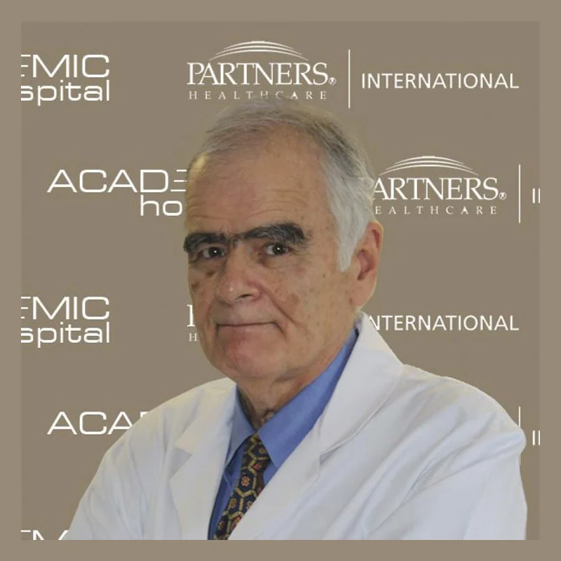
Prof. Dr. Hasan Yazıcı
RheumatologyVerified
- 52+years exp.
English · Turkish
FromPrice on request
Free quote
60 seconds · no obligationView Profile
Calcific tendinitis is a disorder marked by the deposition of calcium hydroxyapatite crystals within tendons, most frequently in the shoulder. It primarily affects working-age adults, causing acute shoulder pain, stiffness, and restricted joint movement that leads patients to seek clinical help. Appropriate therapeutic or procedural options depend on an accurate imaging review and assessment by a rheumatology specialist. Compare hospitals in Turkey that treat Calcific Tendinitis and get a free review of your medical records.

1 doctor Compare doctors, ratings and prices — ask any of them for a free quote.
RheumatologyVerified
English · Turkish
60 seconds · no obligationView Profile
Calcific tendinitis is a disorder marked by the deposition of calcium hydroxyapatite crystals within tendons, most frequently in the shoulder. It primarily affects working-age adults, causing acute shoulder pain, stiffness, and restricted joint movement that leads patients to seek clinical help. Appropriate therapeutic or procedural options depend on an accurate imaging review and assessment by a rheumatology specialist.
Specialty: Rheumatology
The cost of treating Calcific Tendinitis depends on the diagnosis and the treatment chosen. Doctors first review your medical records — free of charge — and you then receive an itemised quote.
Calcific Tendinitis is treated by Rheumatology specialists. 1 clinic in Turkey on our platform offer this treatment.
Calcific tendinitis is a painful disorder characterised by calcium hydroxyapatite deposition within tendon tissue. It most frequently targets the rotator cuff of the shoulder, particularly the supraspinatus tendon, though the infraspinatus and subscapularis can also be involved. The condition primarily affects adults between 30 and 60 years of age, occurring more often in women and people with metabolic disorders such as diabetes or thyroid disease.
Unlike gradual degenerative tendon wear, calcific tendinitis represents an active cellular process with distinct phases. During the formative phase, tendon cells transform into chondrocytes that produce matrix vesicles, gradually forming calcium deposits. This state often remains quiet during a resting period. However, during the subsequent resorptive phase, vascular tissue proliferates around the deposit as the immune system attempts to clear the mineral. This vascular expansion increases internal tendon pressure and causes marked bursal inflammation.
Symptoms often manifest as sudden, intense shoulder pain that wakes patients at night. Elevating the arm, reaching behind the back, or sleeping on the affected side can become almost impossible. Patients frequently experience joint stiffness, mechanical catching, and reduced functional movement. Discomfort during the resorptive phase is often severe enough that individuals seek urgent clinical intervention.
A precise diagnosis combines clinical assessment with targeted diagnostic imaging. When attending a clinic in Turkey, your examination typically begins with an evaluation by a specialist in rheumatology or musculoskeletal medicine. The clinician assesses localized tenderness near the greater tuberosity, tests rotator cuff strength, and evaluates range of motion.
Diagnostic evaluations coordinated through hospital check-ups and diagnostics departments generally include:
To facilitate assessment in Turkey, international patients should bring prior imaging discs in DICOM format, radiology reports, records of past injections or therapy, and a current medication list.
Care plans aim to alleviate pain, relieve mechanical impingement, and restore shoulder mobility. Recommended therapies depend on symptom intensity, deposit texture, and the doctor's review of imaging findings.
| Option | Typically used when | Things to know |
|---|---|---|
| Conservative care | Symptoms are mild, or deposits are in the early formative stage. | Combines oral anti-inflammatory drugs, activity modification, and structured physical therapy to maintain joint motion. |
| Subacromial injection | Acute bursitis or intense pain impairs sleep and rehabilitation. | Delivers corticosteroids and local anaesthetic to reduce inflammation; does not dissolve mineral deposits directly. |
| Ultrasound-guided barbotage | Moderate to severe symptoms persist with visible, accessible deposits. | Uses saline irrigation under local anaesthesia to puncture and aspirate softened calcium paste. |
| Extracorporeal shockwave therapy | Chronic deposits fail conservative care and surgery is deferred. | Acoustic sound pulses stimulate tissue turnover and natural calcium resorption over several sessions. |
| Arthroscopic surgery | Severe symptoms persist after non-operative therapies, or deposits are hard. | Performed by specialists in orthopaedics; uses keyhole instruments to remove deposits and decompress subacromial space. |
Following procedural treatment, guided rehabilitation is essential to restore rotator cuff strength and movement. Certain facilities also discuss supportive methods in regenerative medicine to facilitate tendon healing.
Travelling for medical care requires organized preparation. International patients generally start by submitting recent shoulder imaging and clinical records for remote evaluation. This enables clinical teams in Turkey to verify your diagnosis and suggest procedural or non-surgical paths before travel.
Expected duration of stay varies according to the chosen procedure:
Before departing Turkey, obtain a thorough medical summary in English, including procedure notes, post-treatment imaging, and medication instructions. Arrange follow-up care with your local doctor or physiotherapist to ensure continuous rehabilitation progress.
Travelling long distances with acute shoulder pain introduces physical strain. You should postpone air travel if you notice red-flag indicators requiring urgent local medical evaluation. Seek prompt local care if you develop:
Individuals with unstable chronic illnesses, uncontrolled cardiovascular conditions, or active infections must postpone elective medical travel until cleared by their local physician.
The precise cause remains uncertain, but calcific tendinitis is linked to local cellular changes rather than excessive dietary calcium intake. Reduced local blood supply, tendon hypoxia, and repetitive micro-trauma can prompt stem cells in the tendon to transform into chondrocytes. These chondrocytes deposit calcium hydroxyapatite crystals. Endocrine factors, including diabetes and thyroid disorders, may also increase individual susceptibility to the condition.
Calcific tendinitis involves mineral crystal accumulation inside an intact or irritated tendon, whereas a rotator cuff tear involves a physical breach or rupture in tendon fibres. While both conditions produce shoulder pain and restricted motion, calcific tendinitis frequently triggers sudden, severe pain during its resorptive stage. Diagnostic ultrasound or MRI scans distinguish between the two by showing whether crystals or actual structural tears are present.
Ultrasound-guided barbotage, or lavage, is a minimally invasive outpatient procedure performed under local anaesthetic without general anaesthesia. A clinician uses real-time ultrasound guidance to position one or two needles directly into the calcific deposit. Warm saline is repeatedly injected and aspirated to dissolve and flush out the softened mineral paste. The procedure typically takes thirty to forty-five minutes, followed by a short rest period before discharge.
Yes, many calcific deposits eventually undergo spontaneous resorption as the immune system clears the hydroxyapatite crystals over several months or years. However, the resorptive phase often induces intense pain, inflammation, and joint stiffness. When conservative observation fails to relieve persistent discomfort, or when symptoms severely interfere with sleep and daily activities, medical procedures such as needling or surgery are evaluated.
Following ultrasound-guided barbotage or needle lavage, patients can usually fly within three to five days once immediate soreness subsides and post-procedure mobility is confirmed. If arthroscopic surgery is performed, surgeons generally recommend staying seven to ten days. This window ensures surgical portal healing, allows initial physiotherapy assessment, and minimises the discomfort and circulatory risks associated with air travel after an operation.
Your request is in!
Your coordinator will contact you — usually within 2 hours, 24/7.
Want a faster, more accurate assessment?
Add photos & medical detailsLeave your number — a coordinator calls you in your language, usually within 15 minutes during office hours.
Thank you!
A coordinator will contact you at the time you chose.
