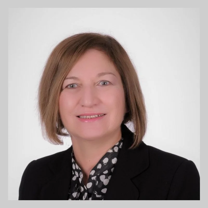
Dr. Tülay Yarkın
Pulmonology & Thoracic SurgeryVerified
- 28+years exp.
English · Turkish
FromPrice on request
Free quote
60 seconds · no obligationView Profile
What a lung biopsy is, how needle, bronchoscopic and surgical biopsy techniques work, what the evidence says about diagnostic accuracy, how much a lung biopsy costs in Turkey compared with the UK, US and Germany, what happens during the procedure, how long recovery takes, possible risks and complications, and how to choose an accredited hospital.
A written plan with the surgeon's name and the full price — usually within 24 hours.

1 doctor Compare doctors, ratings and prices — ask any of them for a free quote.
Pulmonology & Thoracic SurgeryVerified
English · Turkish
60 seconds · no obligationView Profile
A lung biopsy is a diagnostic medical procedure in which a small sample of lung tissue or cells is removed for examination under a microscope. Pathologists analyse the sample to identify abnormal cells, differentiate between benign and malignant tumours, and diagnose chronic inflammatory or interstitial conditions. When a chest X-ray or CT scan reveals an unexplained pulmonary nodule, mass, or persistent shadow, imaging alone cannot establish the underlying diagnosis. A biopsy provides the cellular details needed to determine an accurate treatment plan.
Specialised hospitals in Turkey evaluate international patients through multidisciplinary teams of interventional pulmonologists, thoracic surgeons, and radiologists. This guide explains how needle, bronchoscopic, and surgical biopsies work, what the evidence says about diagnostic accuracy, approximate costs compared with the UK, US, and Germany, recovery milestones, and safety standards. For the wider choice of respiratory care, see our guide to pulmonology treatments in Turkey.
A lung biopsy is performed using one of three primary approaches, chosen according to the size, depth, and anatomical position of the abnormal tissue.
A CT-guided percutaneous needle biopsy targets nodules located in outer regions of the lung near the chest wall. Under local anaesthesia and real-time CT imaging, a specialist guides a fine needle through the chest wall into the nodule to withdraw cells (fine-needle aspiration) or cylindrical tissue cores.
A bronchoscopic biopsy accesses central lesions located near major airways. A flexible bronchoscope is guided through the mouth or nose under sedation. Clinicians often use endobronchial ultrasound (EBUS-TBNA) or navigational bronchoscopy to direct a needle through the airway wall into adjacent lymph nodes or tissue with high precision.
A surgical biopsy, usually video-assisted thoracoscopic surgery (VATS), is used when lesions cannot be reached safely by needle or bronchoscope, or when larger tissue architecture is required. Under general anaesthesia, a thoracic surgeon uses two or three small chest incisions to insert a camera and instruments to excise a wedge of lung tissue.
Clinical evidence confirms that lung biopsies offer high diagnostic accuracy when the appropriate technique is matched to the lesion. For solid peripheral nodules, CT-guided core needle biopsy achieves diagnostic sensitivity between 88% and 95% for malignant disease. When an adequate sample is retrieved, pathologists can identify specific cancer subtypes and perform molecular biomarker testing for targeted therapies.
However, non-diagnostic samples occur in approximately 5% to 15% of needle biopsies. This happens if the needle encounters necrotic tissue, extensive fibrosis, or if small nodules move during breathing. Many Turkish centres utilize rapid on-site evaluation (ROSE), where a cytopathologist checks sample adequacy in the procedure room to reduce the need for repeat procedures.
EBUS-TBNA achieves a diagnostic sensitivity exceeding 90% for mediastinal lymph node staging without chest wall puncture. Surgical VATS biopsy remains the benchmark, delivering a definitive diagnosis in over 98% of cases because intact tissue sections are removed. Trustworthy clinics never guarantee a 100% definitive result from a needle sample alone; if findings remain inconclusive, repeat sampling or surgical excision is advised.
The figures below reflect approximate cost ranges across private hospitals. These estimates represent broad international comparisons rather than fixed quotations, as final charges depend on the technique used, anaesthesia, hospital stay, and laboratory work.
| Country | Approximate cost range |
|---|---|
| Turkey | €1,200 – €4,800 |
| United Kingdom | €2,800 – €8,500 |
| United States | €4,500 – €14,000 |
| Germany | €3,000 – €9,000 |
What changes the price: an outpatient CT needle biopsy under local anaesthesia costs substantially less than a surgical VATS procedure requiring an operating theatre, general anaesthesia, a chest tube, and a hospital stay. Routine histology is also less costly than advanced immunohistochemical (IHC) staining and molecular genetic analysis (such as EGFR, ALK, and PD-L1 testing).
In Turkey, standard international packages include pre-procedure blood tests, imaging review, procedure and doctor fees, anaesthesia or sedation, post-procedure observation, initial pathology reporting, and airport transfers. You can request a clear, itemised free quote before making travel plans.
Choosing the correct biopsy method depends on lesion location and characteristics, balanced against patient safety. Major respiratory guidelines from the British Thoracic Society (BTS) and the American College of Chest Physicians (CHEST) outline clear pathways for each technique.
| Biopsy technique | Best suited to | Typical limits |
|---|---|---|
| CT-guided needle biopsy | Peripheral nodules, subpleural lesions, chest wall masses | Higher pneumothorax risk; small sample volume |
| Bronchoscopy and EBUS | Central airway tumours, mediastinal and hilar lymph nodes | Cannot reliably reach far peripheral nodules without navigational guidance |
| Surgical biopsy (VATS) | Indeterminate nodules, subpleural wedges, diffuse lung disease | Requires general anaesthesia, hospital admission, and a chest drain |
For outer lung nodules near the ribs, CT needle biopsy avoids general anaesthesia. For abnormalities adjacent to central airways or mediastinal lymph nodes, bronchoscopy is preferred because it avoids puncturing the outer pleura. Interventional endoscopy also manages structural airway defects; for example, see our guide to bronchial fistula closure via bronchoscopy. When needle samples are inconclusive, VATS provides direct visual access and conclusive tissue samples.
A lung biopsy is indicated for patients with unexplained pulmonary nodules measuring over 8 millimetres that show suspicious features on CT or PET scans, such as spiculated borders, documented growth, or high metabolic activity. It is also required for staging known cancers, identifying targetable genetic mutations, or diagnosing unexplained interstitial lung fibrosis.
A biopsy is not suitable for every case. Very small nodules under 6 millimetres with low malignant risk are typically monitored with serial low-dose CT scans rather than invasive sampling. Severe uncorrected bleeding disorders represent a contraindication until blood parameters normalize. Patients with severe bullous emphysema face a heightened risk of pneumothorax during needle passage, making bronchoscopic alternatives preferable. Severe pulmonary hypertension or uncompensated respiratory failure also requires medical stabilization before elective biopsy.
A standard hospital biopsy follows five clear clinical steps designed to ensure accuracy and patient comfort.
First, pre-procedure checks take place: fasting for six hours, pausing anticoagulant medications under medical supervision, and placing an intravenous cannula. Second, you are positioned on the CT couch or endoscopy table. For a needle biopsy, local anaesthetic is injected into the skin, chest wall muscles, and pleura to numb the pathway thoroughly.
Third, the specialist introduces the needle or bronchoscope under image guidance. During needle advancement, you are asked to hold your breath briefly to stabilize lung movement. Fourth, tissue specimens are collected: core samples are taken with a biopsy device, or fine-needle aspirates are collected through the bronchoscope. Finally, the instruments are withdrawn, pressure is applied to the puncture site, and a sterile dressing is placed. If VATS was performed, small portal incisions are sutured and a temporary intercostal drain is connected.
Most international patients travelling for a needle biopsy or bronchoscopy stay in Turkey for 3 to 4 nights. Patients undergoing surgical VATS should plan for 5 to 7 nights.
Following a needle biopsy, you rest in an observation ward for two to four hours while medical staff monitor your vital signs. A post-procedure chest X-ray is taken within one to four hours to check for air leakage. Needle and bronchoscopy procedures are usually performed as day cases, allowing return to your hotel the same day. VATS patients stay in hospital for one to two nights until the chest drain is removed.
Over the next two days, rest and gentle walking are encouraged, while vigorous exercise and heavy lifting must be avoided. Reviewing how it works helps international visitors organize airport transfers and clinic appointments smoothly.
Crucially, you must not fly home immediately after a lung biopsy. Cabin pressure changes during flight can cause a small air leak to enlarge into a serious pneumothorax. Thoracic doctors require a follow-up chest X-ray confirming the lung is fully expanded before issuing a fit-to-fly certificate.
Every invasive diagnostic procedure carries potential risks. Clinical data from the British Thoracic Society and the NHS document the main recognized complications.
Pneumothorax (air entering the pleural space around the lung) is the most frequent complication of CT-guided needle biopsy, occurring in 15% to 25% of cases. Most air leaks are minor and heal on their own without intervention. In about 3% to 7% of cases, a larger air leak requires a temporary chest drain to allow the lung to re-expand. Bronchoscopic procedures have a much lower pneumothorax rate, typically under 2%.
Haemoptysis (coughing up minor traces of blood) occurs in 5% to 10% of cases due to localized irritation of bronchial vessels. This almost always stops within 24 to 48 hours. Significant bleeding is rare, seen in fewer than 1% of patients.
Other uncommon complications include puncture site soreness, minor infection, and transient pleural pain, which are managed with simple painkillers. Systemic air embolism is an extremely rare complication (<0.1%). Accredited Turkish hospitals maintain immediate thoracic surgical support and intensive care units to manage any complication safely.
When selecting a hospital for a diagnostic lung procedure, confirm that the facility is licensed by the Turkish Ministry of Health and accredited by international organisations such as the JCI. Ensure the procedure takes place in a fully equipped general hospital with interventional radiology suites, thoracic operating theatres, and an intensive care unit, rather than an unaccredited outpatient clinic.
Check that the hospital has an accredited on-site pathology laboratory capable of rapid on-site evaluation, immunohistochemistry, and molecular tumour testing. Red flags include clinics that guarantee a 100% conclusive needle biopsy without reviewing your scans, coordinators who offer fixed pricing without examining your CT DICOM discs, and facilities that lack emergency thoracic surgery cover. Read our overview of thoracic surgery in Turkey and request a written free quote before confirming your trip.
Before confirming your biopsy appointment, ask your medical coordinator these essential questions:
A lung biopsy is a diagnostic medical procedure where a small sample of lung tissue or cells is removed and analysed under a microscope. It is performed to investigate abnormal pulmonary nodules, persistent lung shadows, masses, or diffuse interstitial lung diseases detected on chest X-rays or CT scans.
The procedure is designed to minimise discomfort. Percutaneous needle biopsies use local anaesthetic to numb the skin, chest wall, and pleura, so you feel pressure rather than sharp pain. Bronchoscopy and EBUS are conducted under conscious sedation or general anaesthesia, and surgical biopsies (VATS) are performed under general anaesthesia.
Diagnostic accuracy is high. CT-guided core needle biopsy achieves diagnostic sensitivity between 88% and 95% for malignant tumours. EBUS-TBNA provides over 90% accuracy for mediastinal lymph node staging. Surgical VATS biopsy offers diagnostic accuracy exceeding 98% because larger, intact tissue sections are removed for histopathological examination.
A lung biopsy in Turkey typically costs between €1,200 and €4,800, compared with €2,800 to €8,500 in the UK and €4,500 to €14,000 in the US. The price varies depending on whether a needle biopsy, bronchoscopy, or surgical VATS procedure is performed, and includes pathology analysis.
A needle biopsy passes through the chest wall from the outside under CT guidance, making it best suited for peripheral nodules near the ribs. A bronchoscopy inserts a thin, lighted tube through your airways from the inside, making it ideal for central airway tumours and mediastinal lymph node sampling.
Recovery depends on the method used. After a CT needle biopsy or bronchoscopy, most patients rest for 24 to 48 hours and resume light daily activities within three days. Surgical VATS biopsies require one to two weeks of reduced activity while the small chest incisions heal.
The most common risk is pneumothorax (air leakage into the pleural cavity), occurring in 15% to 25% of CT-guided needle procedures, though only 3% to 7% need a temporary chest tube. Other risks include minor coughing up of blood (haemoptysis), localized soreness, and rarely infection or bleeding.
Preliminary cytology or rapid on-site evaluation results are often available within 24 to 48 hours. Comprehensive histopathology, including specialized immunohistochemical stains and molecular genetic profiling (such as EGFR, ALK, or PD-L1 markers), typically takes three to five working days to finalize.
For a needle biopsy or bronchoscopy, an itinerary of three to four nights is standard. This allows time for pre-procedure tests, the biopsy, a mandatory post-procedure chest X-ray, and initial results. Surgical VATS biopsies usually require five to seven nights to monitor healing before flight departure.
No. Air travel immediately after a lung biopsy is unsafe because cabin pressure changes can worsen an undetected air leak. Thoracic doctors require a follow-up chest X-ray at least 24 to 48 hours post-procedure to confirm the lung is fully expanded before issuing a fit-to-fly certificate.
Your request is in!
Your coordinator will contact you — usually within 2 hours, 24/7.
Want a faster, more accurate assessment?
Add photos & medical detailsLeave your number — a coordinator calls you in your language, usually within 15 minutes during office hours.
Thank you!
A coordinator will contact you at the time you chose.

