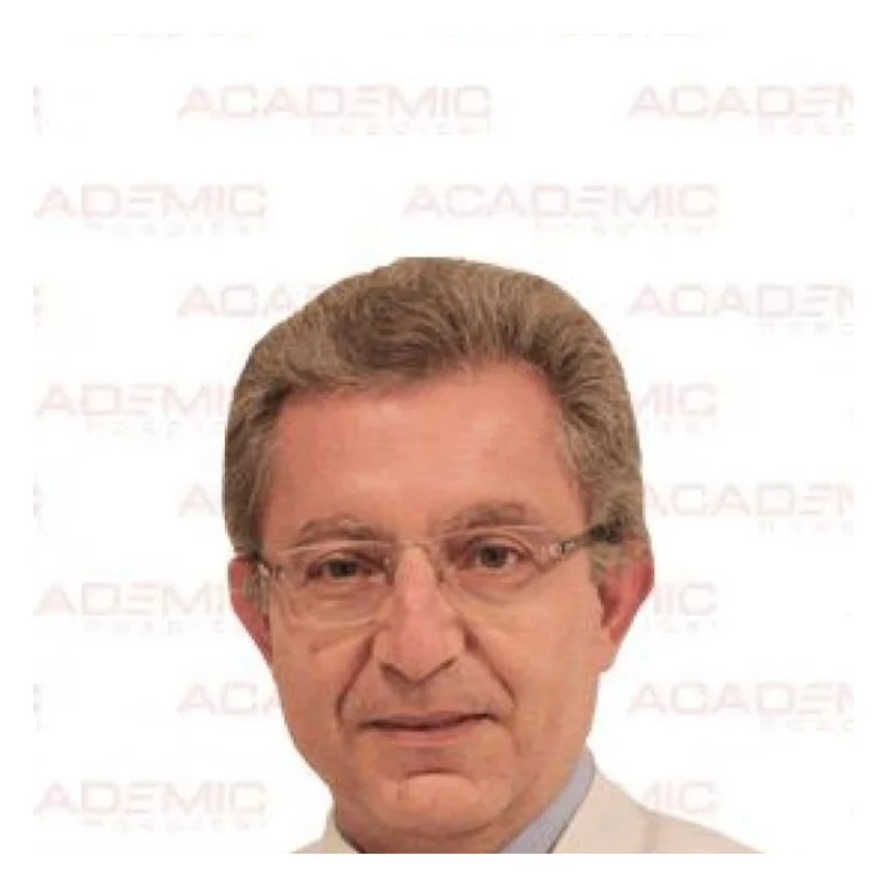
Prof. Dr. Ali Serdar Fak
Cardiac SurgeryVerified
- 29+years exp.
English · Turkish
FromPrice on request
Free quote
60 seconds · no obligationView Profile
What a heart ultrasound is, how sound waves produce moving pictures of the chambers and valves, what a standard, stress and transoesophageal scan each show, what the test cannot detect, approximate costs in Turkey against the UK, US and Germany, preparation, what the scan feels like, how long it takes, how to read a report and when other tests are added.
A written plan with the surgeon's name and the full price — usually within 24 hours.

2 doctors Compare doctors, ratings and prices — ask any of them for a free quote.
Cardiac SurgeryVerified
English · Turkish
60 seconds · no obligationView Profile
Cardiac SurgeryVerified
English · Turkish
60 seconds · no obligationView Profile
A heart ultrasound, also called an echocardiogram or echo, is a scan that uses high-frequency sound waves to produce live moving pictures of the heart. A small handheld probe, coated with gel, is placed on the chest. The sound waves bounce off the heart walls and valves, and a computer turns the echoes into an image that shows how big the chambers are, how thick the muscle is, how well the heart squeezes and how blood flows through the valves. Doppler settings colour the blood flow, so leaks and narrowings can be measured. There is no radiation, no needle and no recovery period.
This guide explains how the scan works, the different types, what it can and cannot show, approximate costs in Turkey against the UK, US and Germany, how to prepare, and how to read the report. For other heart tests and treatments see our cardiology treatments in Turkey.
Doctors request a heart ultrasound to look for the cause of breathlessness, swollen ankles, chest pain, palpitations, fainting or a heart murmur; to check heart function after a heart attack or during chemotherapy; to assess a known valve problem over time; and as part of a general cardiac check-up for people with high blood pressure, diabetes or a family history of heart disease. It is also routine before many operations. Because it is quick and safe, it is often the first imaging test after an ECG shows something unusual.
The scan is well supported for measuring the pumping strength of the left ventricle (ejection fraction), detecting valve narrowing or leakage, spotting fluid around the heart, finding some congenital defects and estimating the pressure in the lung circulation. It shows the heart as it moves, which makes it more informative than a still picture for function.
It has limits. A standard scan cannot reliably show narrowed coronary arteries, and a normal result at rest does not rule out coronary disease. It also cannot capture rhythm problems that come and go, which need an ECG or a longer recording. Image quality depends on body build, lung gas and the operator, and a small number of scans are too unclear to answer the question, in which case a contrast agent or another test is used.
There are several versions, and the name on your request matters because the cost and preparation differ.
| Type | Suits | Typical limits |
|---|---|---|
| Transthoracic (standard) echo | First look at chambers, muscle and valves | Images through the chest wall can be limited |
| Stress echo | Looking for reduced blood flow during effort or a drug | Needs exercise or medicine; takes longer |
| Transoesophageal echo (TOE) | Clear view of valves and clots behind the heart | Needs sedation and a probe swallowed |
| Contrast echo | Poor images or the heart edge needs outlining | Needs a vein line and a contrast injection |
Our guide to echocardiography covers the methods in more detail. If your doctor suspects blocked arteries, a stress test or exercise test or a CT coronary angiogram answers a question that ultrasound cannot.
A scan is most useful when there is a reason: symptoms, a murmur, an abnormal ECG, a family history of cardiomyopathy, high blood pressure that has lasted years, or a treatment such as chemotherapy that can affect the heart. In a person with no symptoms and no risk factors, a routine scan usually finds nothing, and guidelines do not recommend screening everyone. A check-up package can still make sense if you are travelling for treatment anyway or want a baseline for later comparison, but it should not replace a conversation about blood pressure, cholesterol and lifestyle, which matter more for most people.
The figures below are approximate ranges for a private scan, not hospital quotes. Prices depend on the type of study and the report.
| Country | Approximate cost range |
|---|---|
| Turkey | €40 – €250 |
| United Kingdom | €150 – €600 |
| United States | €500 – €3,000 |
| Germany | €100 – €400 |
What changes the price: standard scan or stress or transoesophageal study, the use of contrast, whether a consultant cardiologist reviews the images and writes the report, and whether it comes bundled with an ECG and consultation in a check-up package. A simple scan is rarely a reason to travel by itself, but it can be added to a wider check-up or a treatment trip.
For a standard scan no preparation is needed: eat and take your usual medicines. For a stress echo, wear comfortable shoes and avoid heavy meals and caffeine for several hours, and ask whether to pause beta-blockers. For a transoesophageal scan you must fast for about six hours and arrange to be accompanied, because of the sedation. During a standard scan you lie on your left side, with chest bare or covered, while the operator moves the probe across the ribs and breastbone and asks you to hold your breath for a few seconds. It feels like light pressure and the gel is cool. A standard scan takes about 20 to 45 minutes; a stress echo about an hour; a transoesophageal study about 30 minutes plus time to recover from sedation.
A report usually lists chamber sizes, wall thickness, ejection fraction and valve findings. A normal left ventricular ejection fraction is typically in the range of about 55 to 70 percent, and a figure below about 40 percent suggests heart failure with reduced pumping, according to professional guidance. Words such as trivial or mild regurgitation often describe findings seen in healthy people and may need no treatment, while moderate or severe narrowing or leaks are followed up. Do not interpret a single number alone; the cardiologist reads it alongside your symptoms and other tests. If an abnormality is found, the usual next steps are repeat scans, tests such as the stress test, or treatment, including valve surgery in advanced cases.
There is no recovery time. You can eat, drive and return to normal activity at once after a standard scan, and the results are often discussed the same day. After a transoesophageal scan you need a few hours for the sedation to wear off and a sore throat is common; you should not drive that day. Stress echo carries the small risks of the exercise or medicine used, such as palpitations or chest discomfort, and is done with a doctor and resuscitation equipment present. Contrast agents rarely cause allergic reactions. The NHS and the British Heart Foundation describe standard ultrasound as safe, with no known harm from the sound waves.
A good scan depends on the person doing it. Check that a cardiologist or an accredited echo technician performs the study and that a cardiologist signs the report, that the equipment is modern and that you receive images and a written report, ideally in English. Red flags include a result handed over with no explanation, no cardiologist involved and a price that changes after you arrive. Read how our process works and request a plan through the free quote page.
A heart ultrasound, or echocardiogram, uses sound waves from a handheld probe on the chest to create live pictures of the heart. It shows the chambers, walls and valves and how blood flows through them, with no radiation and no needle.
A standard scan takes about 20 to 45 minutes. A stress echo takes about an hour, and a transoesophageal study takes around 30 minutes plus time to recover from the sedation. You can usually go home straight afterwards.
An approximate range is €40 to €250 for a private scan, against about €150 to €600 in the UK. The price depends on the type of echo, contrast, cardiologist reporting and whether it is part of a check-up package.
No. You feel light pressure from the probe and the gel is cool. A transoesophageal scan needs sedation and a swallowed probe, which can leave a sore throat for a day, but the standard scan involves no needle. Tell the operator if the probe is uncomfortable, as the pressure can be eased.
It measures pumping strength, valve leaks and narrowing, chamber size, wall thickness, fluid around the heart and some congenital defects. It cannot reliably show blocked coronary arteries or intermittent rhythm problems. Further tests are added when the scan raises a question that it cannot answer on its own.
No. An ECG records the heart's electrical activity, while an ultrasound shows its structure and movement. They answer different questions and are often done together as part of a heart check. Ask your doctor which of the two answers your question.
A standard scan needs no preparation. For a stress echo avoid heavy meals and caffeine for several hours, and for a transoesophageal scan you must fast for about six hours and arrange for someone to accompany you.
Standard ultrasound is considered safe and uses no radiation. Stress echo has small risks from the exercise or medicine, and contrast agents rarely cause allergic reactions, so a doctor is present for those studies. Ask the clinic about any allergies before contrast is used.
One day is enough for a standard scan, often with the report the same day. If it is part of a wider check-up, plan one to two days, longer if further tests or treatment are advised.
Your request is in!
Your coordinator will contact you — usually within 2 hours, 24/7.
Want a faster, more accurate assessment?
Add photos & medical detailsLeave your number — a coordinator calls you in your language, usually within 15 minutes during office hours.
Thank you!
A coordinator will contact you at the time you chose.